《山东大学学报(理学版)》 ›› 2020, Vol. 55 ›› Issue (9): 62-71.doi: 10.6040/j.issn.1671-9352.0.2019.475
银温社,贺建峰*( )
)
Wen-she YIN,Jian-feng HE*()
摘要:
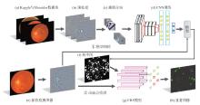
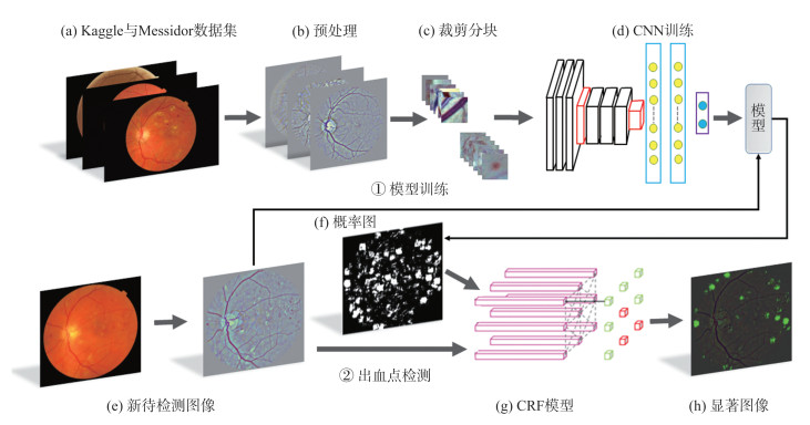
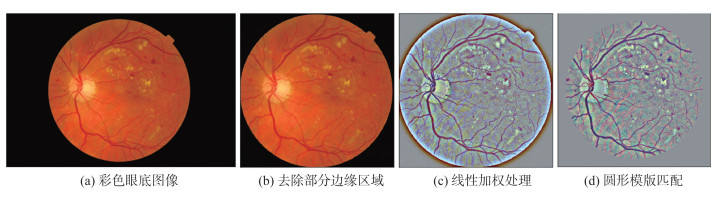
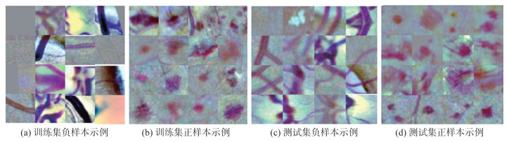
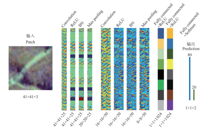
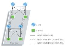
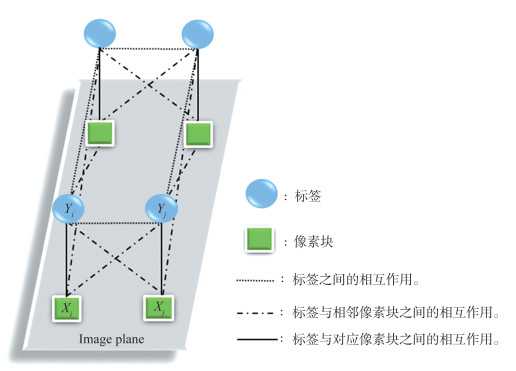
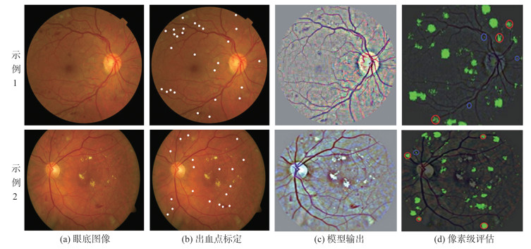
提出了一种基于卷积神经网络(convolutional neural networks,CNN)加条件随机场(conditional random fields,CRF)的眼底图像出血点检测方法。首先,为了避免图像背景区域对后续检测的影响,参考眼底图像中的灰度信息并根据眼底中心位置到其边缘的长度,将图像调整到合适的尺寸,再对图像进行线性加权增强其亮度和对比度;然后,用裁剪到的图像块在仿照VGG网络构建的CNN架构上去训练检测出血点的CNN模型;最后,为了克服CNN模型在出血点检测中误检、漏检等问题,采用CRF对CNN模型输出的概率图进行后处理,以实现眼底图像出血点的精确检测。提出的检测方法在公开的Kaggle与Messidor数据库上进行训练和验证,获得了98.8%的准确率、99.4%的召回率和99.1%的F-score。另外,在DIARETDB1数据库上测试的灵敏度达到98.5%,F-score为96.1%。实验结果表明,从图像视觉和定量检测2个方面均说明了提出方法的有效性和优越性。
中图分类号:
| 1 | GIRARD F. Simultaneous macula detection and optic disc boundary segmentation in retinal fundus images[C]//SPIE Medical Imaging. San Diego: SPIE, 2016. |
| 2 |
SAFI H , SAFI S , HAFEZIMOGHADAM A , et al. Early detection of diabetic retinopathy[J]. Survey of Ophthalmology, 2018, 63 (5): 601- 608.
doi: 10.1016/j.survophthal.2018.04.003 |
| 3 | MA Xiaolong, XIE Xudong, LAM K, et al. A new bottom-up method for saliency detection[C]//IEEE International Symposium on Consumer Electronics. Hsinchu: IEEE, 2013. |
| 4 | 肖志涛, 赵北方, 张芳, 等. 基于k均值聚类和自适应模板匹配的眼底出血点检测方法[J]. 中国生物医学工程学报, 2015, 34 (3): 264- 271. |
| XIAO Zhitao , ZHAO Beifang , ZHANG Fang , et al. Method for detecting fundus hemorrhage point based on k-means clustering and adaptive template matching[J]. Chinese Journal of Biomedical Engineering, 2015, 34 (3): 264- 271. | |
| 5 | HALOI M , DANDAPAT S , SINHA R . A Gaussian scale space approach for exudates detection, classification and severity prediction[J]. Computer Science, 2015, 56 (1): 3- 6. |
| 6 | SRIVASTAVA R , WONG D W , DUAN L , et al. Red lesion detection in retinal fundus images using Frangi-based filters[J]. IEEE Engineering in Medicine and Biology Society, 2015, 2015 (1): 5663- 5666. |
| 7 | YANG Chuan , ZHANG Lihe , LU Huchuan , et al. Saliency detection via graph-based manifold ranking[J]. Computer Vision & Pattern Recognition, 2013, 9 (4): 3166- 3173. |
| 8 | JIANG Huaizu , WANG Jingdong , YUAN Zejian , et al. Salient object detection: a discriminative regional feature integration approach[J]. International Journal of Computer Vision, 2017, 123 (2): 251- 268. |
| 9 |
BORJI A , CHENG M M , JIANG H , et al. Salient object detection: a benchmark[J]. IEEE Transactions on Image Processing, 2015, 24 (12): 5706- 5722.
doi: 10.1109/TIP.2015.2487833 |
| 10 | PRATT H , COENEN F , BROADBENT D M , et al. Convolutional neural networks for diabetic retinopathy[J]. Procedia Computer Science, 2016, 90 (7): 200- 205. |
| 11 |
VAN G M , VAN G B , HOYNG C , et al. Fast convolutional neural network training using selective data sampling:application to hemorrhage detection in color fundus images[J]. IEEE Transactions on Medical Imaging, 2016, 35 (5): 1273- 1284.
doi: 10.1109/TMI.2016.2526689 |
| 12 | YANG Yehui , LI Tao , LI Wensi , et al. Lesion detection and grading of diabetic retinopathy via two-stages deep convolutional neural networks[J]. Medical Image Computing and Computer-Assisted Intervention, 2017, 10435 (3): 533- 540. |
| 13 | LAM C , YU C , HUANG L , et al. Retinal lesion detection with deep learning using image patches[J]. Investigative Ophthalmology & Visual Science, 2018, 59 (1): 590- 596. |
| 14 | RAMON P , SANDRA A , JACQUES W , et al. A data-driven approach to referable diabetic retinopathy detection[J]. Artificial Intelligence in Medicine, 2019, 96 (3): 93- 106. |
| 15 | ORLANDO J I , PROKOFYEVA E , DEL F M , et al. An ensemble deep learning based approach for red lesion detection in fundus images[J]. Computer Methods Programs Biomed, 2018, 153 (10): 115- 127. |
| 16 | 马文婷.面向眼科医学图像的病变检测研究[D].北京:北京交通大学, 2018. |
| MA Wenting. Research on lesion detection for ophthalmic medical images[D]. Beijing: Beijing Jiaotong University, 2018. | |
| 17 | 张诗浩.基于深度学习的眼底图像出血点分割方法研究[D].天津:天津工业大学, 2019. |
| ZHANG Shihao. Research on segmentation method of hemorrhages of fundus image based on deep learning[D]. Tianjin: Tianjin Polytechnic University, 2019. | |
| 18 | GU J , WANG Z , KUEN J , et al. Recent advances in convolutional neural networks[J]. Pattern Recognition, 2018, 77 (1): 354- 377. |
| 19 | UCHIDA K, TANAKA M, OKUTOMI M. Coupled convolution layer for convolutional neural network[C]//International Conference on Pattern Recognition. Cancun: ICPR, 2016. |
| 20 | DECENCI RE E , ZHANG X , CAZUGUEL G , et al. Feedback on a publicly distributed image database: the Messidor database[J]. Image Analysis & Stereology, 2014, 33 (3): 231- 234. |
| 21 | KAUPPI T, KALESNYKIENE V, KAMARAINEN J K, et al. DIARETDB1 diabetic retinopathy database and evaluation protocol[C]//Proceeding of the British Machine Vision Conference. Coventry: DPLP, 2007: 1-10. |
| 22 | DJEKOUNE A O , MESSAOUDI K , AMARA K . Incremental circle hough transform: an improved method for circle detection[J]. Optik - International Journal for Light and Electron Optics, 2017, 133 (1): 17- 31. |
| 23 | 杨俊俐, 姜志国, 周全, 等. 基于条件随机场的遥感图像语义标注[J]. 航空学报, 2015, 36 (9): 3069- 3081. |
| YANG Junli , JIANG Zhiguo , ZHOU Quan , et al. Semantic annotation of remote sensing image based on conditional random field[J]. Journal of Aviation, 2015, 36 (9): 3069- 3081. | |
| 24 | KARIMAGHALOO Z , ARNOLD D L , ARBEL T . Adaptive multi-level conditional random fields for detection and segmentation of small enhanced pathology in medical images[J]. Medical Image Analysis, 2015, 27 (2): 17- 30. |
| 25 | 常亮, 邓小明, 周明全, 等. 图像理解中的卷积神经网络[J]. 自动化学报, 2016, 42 (9): 1300- 1312. |
| CHANG Liang , DENG Xiaoming , ZHOU Mingquan , et al. Convolutional neural network in image understanding[J]. Journal of Automation, 2016, 42 (9): 1300- 1312. | |
| 26 | YAN Hua , HU Tian . Depth estimation with convolutional conditional random field network[J]. Neurocomputing, 2016, 214 (19): 546- 554. |
| 27 | 李宗民, 徐希云, 刘玉杰, 等. 条件随机场像素建模与深度特征融合的目标区域分割算法[J]. 计算机辅助设计与图形学学报, 2018, 30 (6): 29- 36. |
| LI Zongmin , XU Xiyun , LIU Yujie , et al. Target region segmentation algorithm based on conditional random field pixel modeling and depth feature fusion[J]. Journal of Computer Aided Design and Graphics, 2018, 30 (6): 29- 36. |
| [1] | 李妮,关焕梅,杨飘,董文永. 基于BERT-IDCNN-CRF的中文命名实体识别方法[J]. 《山东大学学报(理学版)》, 2020, 55(1): 102-109. |
| [2] | 王文卿,撖奥洋,于立涛,张智晟. 自编码器与PSOA-CNN结合的短期负荷预测模型[J]. 《山东大学学报(理学版)》, 2019, 54(7): 50-56. |
| [3] | 张芳芳,曹兴超. 基于字面和语义相关性匹配的智能篇章排序[J]. 山东大学学报(理学版), 2018, 53(3): 46-53. |
| [4] | 秦静,林鸿飞,徐博. 基于示例语义的音乐检索模型[J]. 山东大学学报(理学版), 2017, 52(6): 40-48. |
| [5] | 何炎祥, 刘健博, 孙松涛, 文卫东. 基于层叠条件随机场的微博商品评论情感分类[J]. 山东大学学报(理学版), 2015, 50(11): 67-73. |
| [6] | 潘清清,周枫,余正涛,郭剑毅,线岩团. 基于条件随机场的越南语命名实体识别方法[J]. 山东大学学报(理学版), 2014, 49(1): 76-79. |
|