JOURNAL OF SHANDONG UNIVERSITY(NATURAL SCIENCE) ›› 2021, Vol. 56 ›› Issue (9): 13-20.doi: 10.6040/j.issn.1671-9352.0.2020.655
Previous Articles Next Articles
San-li YI1,2( ),Jian-ting CHEN1,Jian-feng HE1,*()
CLC Number:
| 1 |
KIRBAS C , QUEK F . A review of vessel extraction techniques and algorithms[J]. ACM Computing Surveys (CSUR), 2004, 36 (2): 81- 121.
doi: 10.1145/1031120.1031121 |
| 2 |
LIU I , SUN Y . Recursive tracking of vascular networks in angiograms based on the detection-deletion scheme[J]. IEEE Transactions on Medical Imaging, 1993, 12 (2): 334- 341.
doi: 10.1109/42.232264 |
| 3 |
CAN A , SHEN H , TURNER J N , et al. Rapid automated tracing and feature extraction from retinal fundus images using direct exploratory algorithms[J]. IEEE Transactions on Information Technology in Biomedicine, 1999, 3 (2): 125- 138.
doi: 10.1109/4233.767088 |
| 4 |
ZANA F , KLEIN J C . Segmentation of vessel-like patterns using mathematical morphology and curvature evaluation[J]. IEEE Transactions on Image Processing, 2001, 10 (7): 1010- 1019.
doi: 10.1109/83.931095 |
| 5 |
MENDONCA A M , CAMPILHO A . Segmentation of retinal blood vessels by combining the detection of centerlines and morphological reconstruction[J]. IEEE Transactions on Medical Imaging, 2006, 25 (9): 1200- 1213.
doi: 10.1109/TMI.2006.879955 |
| 6 |
CHAUDHURI S , CHATTERJEE S , KATZ N , et al. Detection of blood vessels in retinal images using two-dimensional matched filters[J]. IEEE Transactions on Medical Imaging, 1989, 8 (3): 263- 269.
doi: 10.1109/42.34715 |
| 7 |
HOOVER A D , KOUZNETSOVA V , GOLDBAUM M . Locating blood vessels in retinal images by piecewise threshold probing of a matched filter response[J]. IEEE Transactions on Medical Imaging, 2000, 19 (3): 203- 210.
doi: 10.1109/42.845178 |
| 8 |
KASS M , WITKIN A , TERZOPOULOS D . Snakes: active contour models[J]. International Journal of Computer Vision, 1988, 1 (4): 321- 331.
doi: 10.1007/BF00133570 |
| 9 | ESPONA L, CARREIRA M J, ORTEGA M, et al. A snake for retinal vessel segmentation[C]//Iberian Conference on Pattern Recognition and Image Analysis. Berlin: Springer, 2007: 178-185. |
| 10 |
STAAL J , ABRMOFF M D , NIEMEIJER M , et al. Ridge-based vessel segmentation in color images of the retina[J]. IEEE Transactions on Medical Imaging, 2004, 23 (4): 501- 509.
doi: 10.1109/TMI.2004.825627 |
| 11 |
RICCI E , PERFETTI R . Retinal blood vessel segmentation using line operators and support vector classification[J]. IEEE Transactions on Medical Imaging, 2007, 26 (10): 1357- 1365.
doi: 10.1109/TMI.2007.898551 |
| 12 | FRAZ M M, REMAGNINO P, HOPPE A, et al. A supervised method for retinal blood vessel segmentation using line strength, multiscale Gabor and morphological features[C]//2011 IEEE International Conference on Signal and Image Processing Applications (ICSIPA). [S. l. ]: IEEE, 2011: 410-415. |
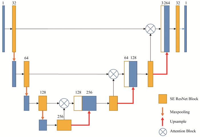
| 13 | RONNEBERGER O, FISCHER P, BROX T. U-net: Convolutional networks for biomedical image segmentation[C]//International Conference on Medical Image Computing and Computer-assisted Intervention. Cham: Springer, 2015: 234-241. |
| 14 | WU Y, XIA Y, SONG Y, et al. Multiscale network followed network model for retinal vessel segmentation[C]//International Conference on Medical Image Computing and Computer-Assisted Intervention. Cham: Springer, 2018: 119-126. |
| 15 | FAN Z, MO J, QIU B, et al. Accurate retinal vessel segmentation via octave convolution neural network[EB/OL]. (2019-06-28)[2020-05-18]. https://arxiv.org/abs/1906.12193v1. |
| 16 |
KOMATSU R , FUJII H , TAMURA Y , et al. Octave deep plane-sweeping network: reducing spatial redundancy for learning-based plane-sweeping stereo[J]. IEEE Access, 2019, 7, 150306- 150317.
doi: 10.1109/ACCESS.2019.2947195 |
| 17 | SZEGEDY C, IOFFE S, VANHOUCKE V, et al. Inception-v4, inception-resnet and the impact of residual connections on learning[C]//Proceedings of the Thirty-first AAAI Conference on Artificial Intelligence. [S. l. ]: AAAI, 2017: 4278-4284 |
| 18 | HU J , SHEN L , SUN G . Squeeze-and-excitation networks[J]. IEEE Transactions on Pattern Analysis and Machine Intelligence, 2018, 42 (8): 2011- 2023. |
| 19 | OKTAY O, SCHLEMPER J, FOLGOC L L, et al. Attention U-net: learning where to look for the pancreas[J/OL]. (2018-04-13)[2020-05-18]. https://arxiv.org/abs/1804.03999v2. |
| 20 | OWEN C G , RUDNICKA A R , MULLEN R , et al. Measuring retinal vessel tortuosity in 10-year-old children: validation of the computer-assisted image analysis of the retina (CAIAR) program[J]. Investigative Ophthalmology & Visual Science, 2009, 50 (5): 2004- 2010. |
| 21 |
VEGA R , SANCHEZ-ANTE G , FALCON-MORALES L E , et al. Retinal vessel extraction using lattice neural networks with dendritic processing[J]. Computers in Biology and Medicine, 2015, 58, 20- 30.
doi: 10.1016/j.compbiomed.2014.12.016 |
| 22 |
LISKOWSKI P , KRAWIEC K . Segmenting retinal blood vessels with deep neural networks[J]. IEEE Transactions on Medical Imaging, 2016, 35 (11): 2369- 2380.
doi: 10.1109/TMI.2016.2546227 |
| 23 | ALOM M Z , YAKOPCIC C , HASAN M , et al. Recurrent residual U-net for medical image segmentation[J]. Journal of Medical Imaging, 2019, 6 (1): 014006. |
| 24 |
ROYCHOWDHURY S , KOOZEKANANI D D , PARHI K K . Iterative vessel segmentation of fundus images[J]. IEEE Transactions on Biomedical Engineering, 2015, 62 (7): 1738- 1749.
doi: 10.1109/TBME.2015.2403295 |
| 25 | LI Q , FENG B , XIE L P , et al. A cross-modality learning approach for vessel segmentation in retinal images[J]. IEEE Transactions on Medical Imaging, 2015, 35 (1): 109- 118. |
| [1] | LIU Zhan-jie1, MA Ru-ning1, ZOU Guo-ping1, ZHONG Bao-jiang2, DING Jun-di 3. An algorithm for color image segmentation based on region growth [J]. J4, 2010, 45(7): 76-80. |
|