《山东大学学报(理学版)》 ›› 2021, Vol. 56 ›› Issue (9): 13-20.doi: 10.6040/j.issn.1671-9352.0.2020.655
易三莉1,2( ),陈建亭1,贺建峰1,*()
San-li YI1,2(),Jian-ting CHEN1,Jian-feng HE1,*()
摘要:
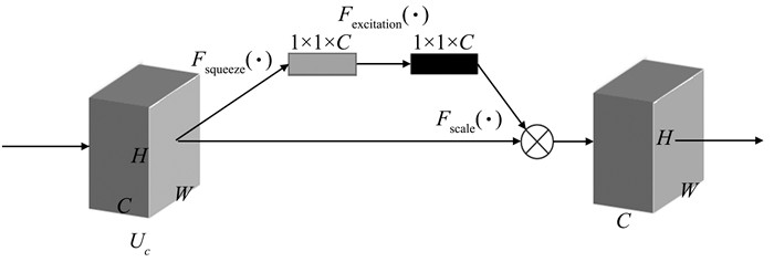
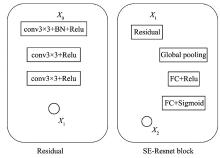
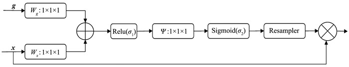
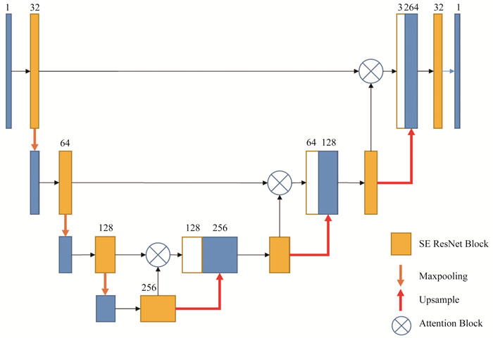
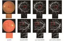
针对现有算法因视网膜图像中血管细小和光照等因素导致的分割精度低的问题, 在U-Net的基础上进行改进, 提出了一种能够较好地提取血管结构的算法模型ASR-UNet。首先, 在编码和解码阶段使用了SE-Resnet结构, 引入通道注意力机制对血管细微结构进行通道增强, 之后在跳跃连接部分使用了AG模块对血管细微结构进行空间增强, 提高网络模型对血管细微结构的分割能力。在公开数据集DRIVE和CHASE_DB1上验证了本文的算法, 在评价指标Acc上分别为0.9697和0.9657, 在敏感性上分别为0.8044和0.7673, 在特异性指标上为0.9859和0.9866。实验结果表明, 近年来的视网膜血管分割算法相比, 本文提出的算法在性能有更好的表现。
中图分类号:
| 1 |
KIRBAS C , QUEK F . A review of vessel extraction techniques and algorithms[J]. ACM Computing Surveys (CSUR), 2004, 36 (2): 81- 121.
doi: 10.1145/1031120.1031121 |
| 2 |
LIU I , SUN Y . Recursive tracking of vascular networks in angiograms based on the detection-deletion scheme[J]. IEEE Transactions on Medical Imaging, 1993, 12 (2): 334- 341.
doi: 10.1109/42.232264 |
| 3 |
CAN A , SHEN H , TURNER J N , et al. Rapid automated tracing and feature extraction from retinal fundus images using direct exploratory algorithms[J]. IEEE Transactions on Information Technology in Biomedicine, 1999, 3 (2): 125- 138.
doi: 10.1109/4233.767088 |
| 4 |
ZANA F , KLEIN J C . Segmentation of vessel-like patterns using mathematical morphology and curvature evaluation[J]. IEEE Transactions on Image Processing, 2001, 10 (7): 1010- 1019.
doi: 10.1109/83.931095 |
| 5 |
MENDONCA A M , CAMPILHO A . Segmentation of retinal blood vessels by combining the detection of centerlines and morphological reconstruction[J]. IEEE Transactions on Medical Imaging, 2006, 25 (9): 1200- 1213.
doi: 10.1109/TMI.2006.879955 |
| 6 |
CHAUDHURI S , CHATTERJEE S , KATZ N , et al. Detection of blood vessels in retinal images using two-dimensional matched filters[J]. IEEE Transactions on Medical Imaging, 1989, 8 (3): 263- 269.
doi: 10.1109/42.34715 |
| 7 |
HOOVER A D , KOUZNETSOVA V , GOLDBAUM M . Locating blood vessels in retinal images by piecewise threshold probing of a matched filter response[J]. IEEE Transactions on Medical Imaging, 2000, 19 (3): 203- 210.
doi: 10.1109/42.845178 |
| 8 |
KASS M , WITKIN A , TERZOPOULOS D . Snakes: active contour models[J]. International Journal of Computer Vision, 1988, 1 (4): 321- 331.
doi: 10.1007/BF00133570 |
| 9 | ESPONA L, CARREIRA M J, ORTEGA M, et al. A snake for retinal vessel segmentation[C]//Iberian Conference on Pattern Recognition and Image Analysis. Berlin: Springer, 2007: 178-185. |
| 10 |
STAAL J , ABRMOFF M D , NIEMEIJER M , et al. Ridge-based vessel segmentation in color images of the retina[J]. IEEE Transactions on Medical Imaging, 2004, 23 (4): 501- 509.
doi: 10.1109/TMI.2004.825627 |
| 11 |
RICCI E , PERFETTI R . Retinal blood vessel segmentation using line operators and support vector classification[J]. IEEE Transactions on Medical Imaging, 2007, 26 (10): 1357- 1365.
doi: 10.1109/TMI.2007.898551 |
| 12 | FRAZ M M, REMAGNINO P, HOPPE A, et al. A supervised method for retinal blood vessel segmentation using line strength, multiscale Gabor and morphological features[C]//2011 IEEE International Conference on Signal and Image Processing Applications (ICSIPA). [S. l. ]: IEEE, 2011: 410-415. |
| 13 | RONNEBERGER O, FISCHER P, BROX T. U-net: Convolutional networks for biomedical image segmentation[C]//International Conference on Medical Image Computing and Computer-assisted Intervention. Cham: Springer, 2015: 234-241. |
| 14 | WU Y, XIA Y, SONG Y, et al. Multiscale network followed network model for retinal vessel segmentation[C]//International Conference on Medical Image Computing and Computer-Assisted Intervention. Cham: Springer, 2018: 119-126. |
| 15 | FAN Z, MO J, QIU B, et al. Accurate retinal vessel segmentation via octave convolution neural network[EB/OL]. (2019-06-28)[2020-05-18]. https://arxiv.org/abs/1906.12193v1. |
| 16 |
KOMATSU R , FUJII H , TAMURA Y , et al. Octave deep plane-sweeping network: reducing spatial redundancy for learning-based plane-sweeping stereo[J]. IEEE Access, 2019, 7, 150306- 150317.
doi: 10.1109/ACCESS.2019.2947195 |
| 17 | SZEGEDY C, IOFFE S, VANHOUCKE V, et al. Inception-v4, inception-resnet and the impact of residual connections on learning[C]//Proceedings of the Thirty-first AAAI Conference on Artificial Intelligence. [S. l. ]: AAAI, 2017: 4278-4284 |
| 18 | HU J , SHEN L , SUN G . Squeeze-and-excitation networks[J]. IEEE Transactions on Pattern Analysis and Machine Intelligence, 2018, 42 (8): 2011- 2023. |
| 19 | OKTAY O, SCHLEMPER J, FOLGOC L L, et al. Attention U-net: learning where to look for the pancreas[J/OL]. (2018-04-13)[2020-05-18]. https://arxiv.org/abs/1804.03999v2. |
| 20 | OWEN C G , RUDNICKA A R , MULLEN R , et al. Measuring retinal vessel tortuosity in 10-year-old children: validation of the computer-assisted image analysis of the retina (CAIAR) program[J]. Investigative Ophthalmology & Visual Science, 2009, 50 (5): 2004- 2010. |
| 21 |
VEGA R , SANCHEZ-ANTE G , FALCON-MORALES L E , et al. Retinal vessel extraction using lattice neural networks with dendritic processing[J]. Computers in Biology and Medicine, 2015, 58, 20- 30.
doi: 10.1016/j.compbiomed.2014.12.016 |
| 22 |
LISKOWSKI P , KRAWIEC K . Segmenting retinal blood vessels with deep neural networks[J]. IEEE Transactions on Medical Imaging, 2016, 35 (11): 2369- 2380.
doi: 10.1109/TMI.2016.2546227 |
| 23 | ALOM M Z , YAKOPCIC C , HASAN M , et al. Recurrent residual U-net for medical image segmentation[J]. Journal of Medical Imaging, 2019, 6 (1): 014006. |
| 24 |
ROYCHOWDHURY S , KOOZEKANANI D D , PARHI K K . Iterative vessel segmentation of fundus images[J]. IEEE Transactions on Biomedical Engineering, 2015, 62 (7): 1738- 1749.
doi: 10.1109/TBME.2015.2403295 |
| 25 | LI Q , FENG B , XIE L P , et al. A cross-modality learning approach for vessel segmentation in retinal images[J]. IEEE Transactions on Medical Imaging, 2015, 35 (1): 109- 118. |
| [1] | 王磊,谢江宁. 基于层级分割的颜色恒常性算法[J]. 山东大学学报(理学版), 2016, 51(1): 101-105. |
| [2] | 刘战杰1,马儒宁1,邹国平1,钟宝江2,丁军娣3. 一种新的基于区域生长的彩色图像分割算法[J]. J4, 2010, 45(7): 76-80. |
| [3] | 徐光柱1,刘鸣2,任东1,马义德3,刘晓丽1. 基于脉冲耦合神经网络的多区域图像分割[J]. J4, 2010, 45(7): 86-93. |
| [4] | 董吉文,杨 森,鲁守银 . 基于单目视觉的移动机器人导航方法[J]. J4, 2008, 43(11): 1-04 . |
|